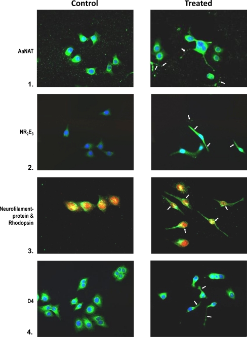
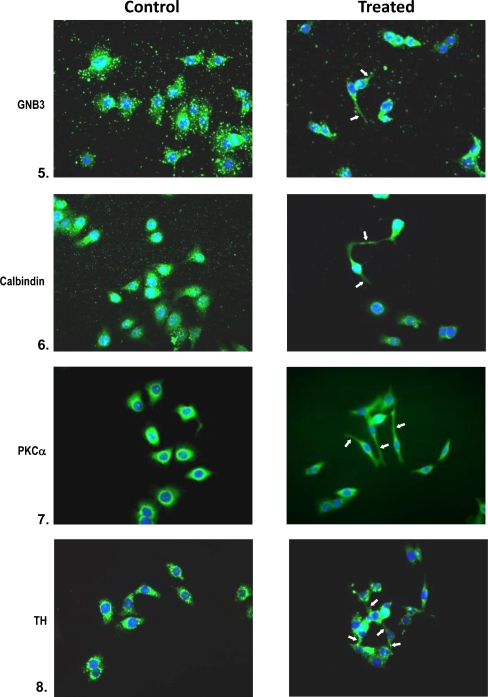
Fig. 6.
Immunophenotyping of retinal progenitors exposed to 50% conditioned medium in low-density cultures on day 2. Controls are untreated cultures. Treated cells were analyzed for the expression of AaNat, Nr2e3, rhodopsin, GNB3, calbindin (rods, cones), PKCα (rod and cone bipolars), and TH (amacrine cells). Note in the composite the neuronal phenotype with extended neurites and axons in treated cultures (arrows). Note the photoreceptor phenotype (1, 2, 3, 4, 5). Note in 3 that photoreceptors are positive for neurofilament protein (red) and rhodopsin (green). Note the bipolars expressing PKCα in 7. In untreated cells, antigens were present but neuronal phenotype was missing


