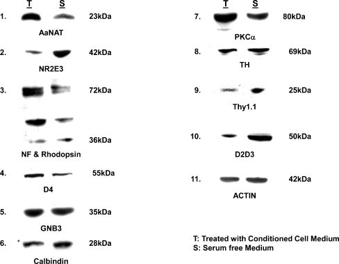
Fig. 7.
Western blot analysis of retinal cells plated at low cell density (1 × 10⁴ cells/cm2) were treated with 50% conditioned medium and assessed by western blot analysis for expression of retinal antigens, day 2. Note the significant upregulation of AaNat and rhodopsin in treated cultures confirming rod photoreceptor differentiation. Both monomeric and dimeric forms of rhodopsin are expressed. However, Nr2e3, a transcription factor for rod photoreceptors, is expressed at higher levels in the controls. A similar increase in treated cultures is seen in the expression of PKCα (expressed in rod and cone bipolars). Also, a slight increase in GNB3, a cone transducin, is noted. No dramatic differences were seen in the expression of other antigens. Actin levels in treated and untreated cultures were similar

