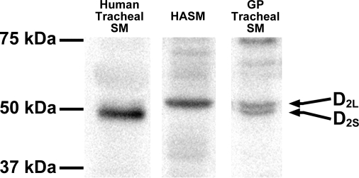
Fig. 2.
Representative immunoblot analyses using antibodies against the dopamine D2 receptor using protein prepared from cultured HASM cells (100 μg), freshly dissected native human tracheal airway smooth muscle (SM; 100 μg), and freshly dissected native guinea pig (GP) tracheal SM (100 μg) identifying two known isoforms of the dopamine D2 receptor (D2L and D2S). White spaces between the 3 lanes indicate that these lanes were located on the same immunoblot but were not located in neighbouring lanes on the original gel and immunoblot image.

