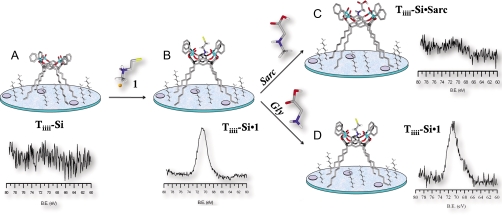
Fig. 4.
XPS analysis of Br 3D region along all steps of the sarcosine recognition protocol in water. (A) pristine Tiiii-Si wafer and its XPS spectrum. (B) Tiiii-Si•1 and its XPS spectrum after exposure of the wafer to a water solution of 1. (C) Tiiii-Si•Sarc and its XPS spectrum after exposure of the wafer to a water solution of sarcosine. (D) Tiiii-Si•1 and its XPS spectrum after exposure of the wafer to a water solution of glycine.

