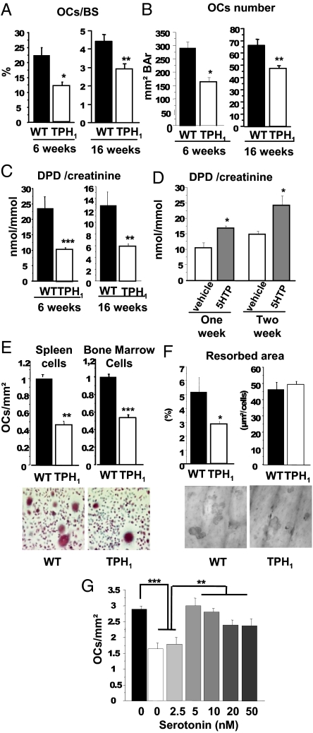
Fig. 2.
Lack of serotonin induces in vivo and in vitro decrease in bone resorption. Histomorphometric analysis was performed at 6 and 16 wk. TRAP staining of a section of the trabecular region of the distal femur was performed to identify osteoclasts. Bone resorption parameters were determined. (A) The number of osteoclasts per bone surface (OCs/BS) showed a marked decrease, and TPH1−/− mice had a lower osteoclast count than WT mice. (B) Osteoclast number/bone area (OCs number/BAr). (C) Biochemical markers of bone resorption. Urinary deoxypyridinoline cross-links normalized by the amount of creatinine present (DPD/creat) were measured in 6- and 16-wk-old WT and TPH1−/− mice. DPD levels were lower in 6- and 16-wk-old male TPH1−/− mice. WT, black bars; TPH1−/−, white bars. Results are reported as mean ± SEM; n = 8 mice per genotype. (D) TPH1−/− mice were treated twice a day with vehicle (white bars) or with 50 mg/kg body weight of 5-HTP (gray bars). DPD levels were significantly increased by 5-HTP administration to 4-wk-old mice after 1 or 2 wk of treatment; vehicle, n = 6; 5-HTP treatment, n = 9. (E) Osteoclastogenesis was assessed in WT and TPH1−/− spleen cells and bone marrow cells cultured with dialyzed serum without serotonin in the presence of M-CSF (25 ng/mL) for 4 d and of M-CSF and RANKL (30 ng/mL) for a further 5 d. Counts of OCLs in WT (black bars) and TPH1−/− (white bars) culture; osteoclast numbers were lower in TPH1−/− cultures than in WT cultures at the end of the differentiation of spleen cells and bone marrow cells. Representative pictures of WT and TPH1−/− spleen cell cultures (20× magnification). (F) OCL activity was assessed by pit assays on dentin slices. The resorbed area was stained with toluidine blue (10× magnification). The resorbed area was strongly decreased in TPH1−/− cultures, whereas the ratio pit area:OC number was unchanged. (G) WT spleen cells were cultured without any serotonin treatment, and TPH1−/− cells were treated with serotonin (2.5, 5, 10, 20, and 50 nM) throughout the culture in the presence of RANKL. From 5 nM 5-HT the number of OCLs increased to equal WT levels, without any dose effect. *P < 0.01 versus WT, **P < 0.001 versus WT, ***P < 0.0001 versus WT.

