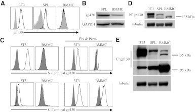
Figure 2. gp130 in resting BMMC is incompletely processed for cell-surface expression.
(A) Primary mouse splenocytes (SPL), the 3T3 fibroblast line, and resting BMMC were examined for gp130 on the cell surface using flow cytometry and (B) gp130 mRNA by RT-PCR. (C) Flow cytometry of BMMC and 3T3 cells using intracellular staining (fixed and permeabilized cells) with gp130 antibodies specific for the N-terminal or the C-terminal compares localization of gp130 protein components. (D) Immunoblot of total cell lysate using gp130 antibodies specific for the N-terminal (N′) and the (E) C-terminal (C') components of the gp130 protein. Shaded histograms (A and C) in flow cytometry data represent isotype control antibody fluorescence.

