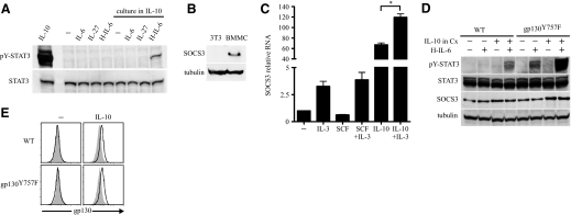
Figure 4. IL-10 permits IL-6 trans-signaling that is negatively regulated by SOCS3.
(A) BMMC were cultured with or without IL-10 (10 ng/ml) for 72 h. Cells washed and rested for 90 min were stimulated with the cytokines shown (each 100 ng/ml) for 15 min and processed for immunoblot. (B) SOCS3 protein was assessed by immunoblot of lysate from resting BMMC cultured in IL-3 and 3T3 cells. (C) BMMC rested for 90 min were stimulated with the cytokines shown (each 10 ng/ml) for 3 h and then total RNA isolated and quantitative PCR performed. RNA samples were pooled from two independent experiments and run under the same PCR conditions. *P < 0.01. (D) WT and gp130Y757F BMMC were cultured with or without IL-10 (10 ng/ml) for 36 h. Cells washed and rested for 90 min were stimulated with H-IL-6 (100 ng/ml) for 15 min where indicated and processed for immunoblot. Data are representative of two experiments with similar results. (E) WT and gp130Y757F BMMC were cultured with or without IL-10 (10 ng/ml) for 36 h and then harvested for flow cytometry. Unless otherwise indicated, all data represent at least three experiments. Shaded histograms in flow cytometry data represent isotype control antibody fluorescence.

