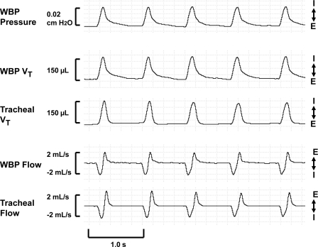
Fig. 2.
Representative recording of a tidal volume (Vt) and airflow validation study recording in a single, anesthetized mouse. WBP Vt was similar in amplitude and waveform morphology to tracheal Vt during the inspiratory limb and demonstrated a gradual roll-off or shoulder before returning to baseline in the expiratory limb. Compared with tracheal airflow, WBP inspiratory flow (I) showed a similar amplitude and morphology, while expiratory flow (E) demonstrated an attenuation in signal amplitude but similar morphology. Signals (from top to bottom) include WBP pressure, WBP Vt, tracheal Vt, WBP airflow, and tracheal airflow.

