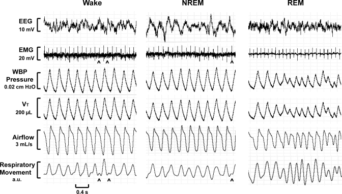
Fig. 6.
Recording sections of a WBP full polysomnographic study demonstrating respiratory waveforms during quiet wakefulness (left), non-rapid eye movement (NREM) sleep (middle), and rapid eye movement (REM) sleep (right) in one mouse. Signals (from top to bottom) include electroencephalographic (EEG) signal, nuchal electromyographic (EMG) signal, WBP chamber pressure, WBP Vt, WBP airflow, and respiratory movement (surrogate for respiratory effort). Intermittent cardiac artifact (carets) can be seen in the EMG and respiratory movement channels.

