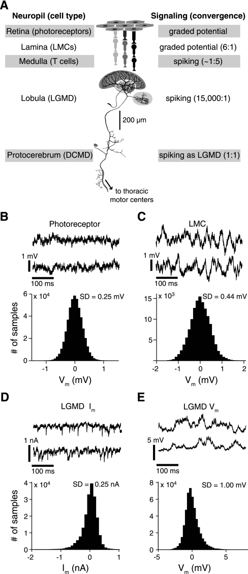
Fig. 1.
Spontaneous variability observed throughout an excitatory visual pathway involved in collision detection. A: schematic illustration of the pathway with its main anatomic and signaling properties. The excitatory input is successively relayed from photoreceptors to large monopolar cells (LMCs), which both respond in a graded manner under our stimulus conditions, and medullary T cells before impinging on the large dendritic fan of the lobula giant movement detector (LGMD; dark gray shading). The 2 smaller dendritic fields receive inhibitory inputs (light gray shading). B, top: two 400-ms long traces illustrating the spontaneous membrane potential (Vm) variability of a typical photoreceptor; bottom: histogram showing the distribution of Vm for the same cell relative to the resting potential. C: spontaneous Vm traces and histogram for a recording from a LMC. D and E: similar traces and histograms of spontaneous membrane currents (Im; voltage clamp) and Vm (current clamp) in the LGMD. SD, standard deviation for the illustrated experiments.

