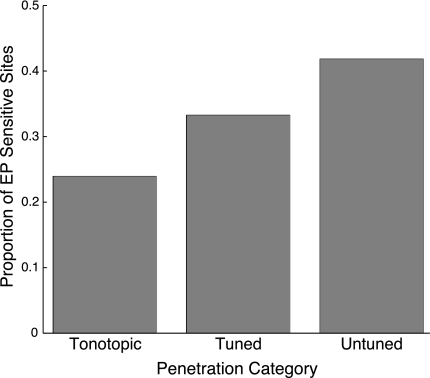
Fig. 3.
Extent of eye position sensitivity by functionally defined region in the inferior colliculus (IC). Bars indicate the proportion of eye position-sensitive recordings within each category. Tonotopic penetrations were those that showed an increase of best frequency as the electrode advanced along a dorsolateral-to-ventromedial trajectory. Frequency-tuned penetrations contained recordings that had well-defined best frequencies but no systematic change with depth. Frequency-untuned penetrations did not show tuning at more than 2 sites. The locations of these penetration categories are indicated in Fig. 4. EP, eye position.

