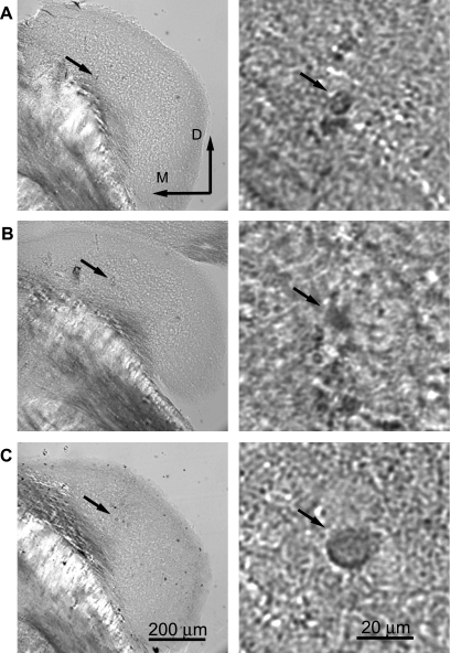
Fig. 1.
Recording sites were marked by iontophoresis of neurobiotin from the recording pipette. Tissue was processed with standard protocols using either horseradish peroxidase (HRP) or avidin Alexa Fluor. Neurons could not be identified unequivocally since the dendrites were not labeled; however, the location in dorsal cochlear nucleus is clear. Right: same tissue at higher magnification. A: neuron that produced a type III-i response [best frequency (BF) = 45 kHz, threshold = 28 dB SPL]. B: type III (BF = 34 kHz, threshold = 13 dB SPL). C: type III (BF = 24 kHz, threshold = 32 dB SPL).

