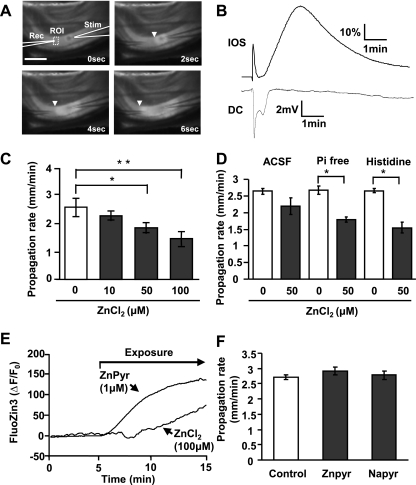
Fig. 1.
Extracellular Zn2+ inhibits spreading depolarization (SD) propagation. A: images showing electrode placement and propagation of SD through the hippocampal CA1 subregion. SD was generated by local microinjection of KCl (10-ms pulse) from a stimulation pipette (Stim) and detected using an extracellular recording electrode (Rec). SD was also tracked from propagating intrinsic optical signal (IOS) increases (>575-nm transmission; arrowheads). Scale bar = 250 μm. ROI, region of interest. B: time courses of IOS and electrical [direct current (DC)] signals generated from the preparation illustrated in A. C: brief ZnCl2 preexposures [10 min in histidine-supplemented artificial cerebrospinal fluid (ACSF)] resulted in concentration-dependent reductions in SD propagation rate (*P < 0.05, **P < 0.01; n = 5). D: comparison of buffer composition on effectiveness of ZnCl2 applications. ZnCl2 (50 μM; 10 min) was more effective in phosphate-free ACSF (Pi-free) and histidine-supplemented ACSF (200 μM histidine) than in control ACSF (*P < 0.05; n = 5). E: intracellular Zn2+ accumulation assessed in single neurons loaded with FluoZin-3. ZnCl2 exposures (100 μM) resulted in slow intracellular accumulation, and more rapid increases were observed with ZnPyr (1 μM ZnCl2 with 100 nM pyrithione). ΔF/F0, relative change in fluorescent intensity. F: preferentially loading intracellular Zn2+ had no effect on SD propagation rate. Exposure to ZnPyr (as in E, followed by brief wash) did not reduce SD propagation rate, and no effects were observed with a control for nonspecific effects of pyrithione (sodium pyrithione, NaPyr; 100 nM; n = 5).

