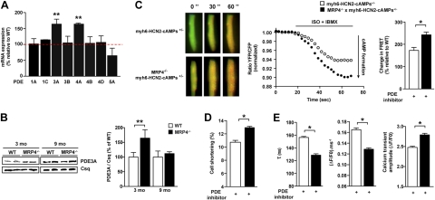
Figure 4.
PDEs are up-regulated and compensate for loss of MRP4 in mice. A) Quantification of mRNA levels of various PDEs by real-time PCR in total cardiac extracts from 3-mo-old mice; n = 5/group in triplicate. B) Quantification of PDE3A protein by Western blot in total cardiac extracts from 3- and 9-mo-old mice (n=5). C) cAMP dynamics and quantification of the changes in cAMP-FRET ratios in cardiacmyocytes from 3-mo-old WT and Mrp4−/− mice after whole-cell stimulation with iso (1 nM) in combination with the nonselective PDE inhibitor IBMX (300 μM). Representative ratiometric images and experiments after iso+IBMX treatment of 7–18 cells (4–6 mice). D, E) Cell shortening (D) and Ca2+ transient parameters (E) of isolated ventricular myocytes from 3-mo-old WT and Mrp4−/− mice measured in the presence of cilostamide (1 μM; 16–24 cells, 3 mice/group). *P < 0.05; **P < 0.001.

