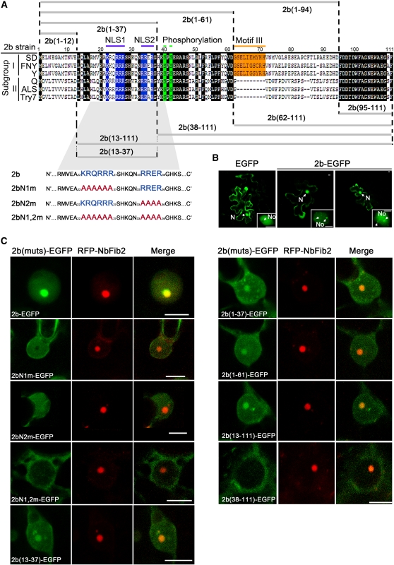
Figure 1.
Subcellular Localization of SD2b and Its Derivative Mutant Proteins.
(A) Alignment of 2b protein sequences from different subgroups of CMV and schematic of SD2b deletion mutants. The two NLS sequences, putative phosphorylation sites, and subgroup I–specific Motif III are highlighted in blue, green, and orange, respectively, and other conserved regions are highlighted in black. Two Arg-rich putative NLSs (blue letters) of the SD2b coding sequence were substituted with Ala residues (red letters) as previously reported (Lucy et al., 2000) and are also shown.
(B) Subcellular localization assays performed by bombarding 35S-SD2b-EGFP into 3-week-old Arabidopsis leaves. The empty EGFP vector was also bombarded as control. One of the typical cells from each bombardment assay for confocal microscopy analysis is presented. The nucleus (N), nucleolus (No), and possible nuclear bodies are labeled with arrows. Bars = 2.5 μm.
(C) Subcellular localization assays performed by coexpression of RFP-NbFib2 with 35S-SD2b-EGFP or mutant proteins fused with EGFP via Agrobacterium-mediated infiltration. One of the typical cells from each assay for confocal microscopy analysis is presented. Bars = 5 μm.

