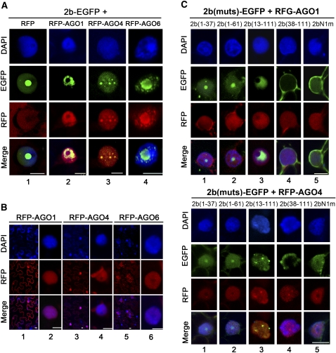
Figure 8.
Colocalization Pattern of SD2b-AGO Complexes in N. benthamiana Leaf Epidermal Cells.
(A) Subcellular colocalization of SD2b-EGFP and RFP-AGO proteins. SD2b-EGFP coinfiltrated with empty RFP vector serves as control. One representative colocalization image is shown for each experiment.
(B) Subcellular location of RFP-AGO1, RFP-AGO4, and RFP-AGO6.
(C) Subcellular location of coexpression of SD2b derivative mutants with RFP-AGO1 (top panel) and RFP-AGO4 (bottom panel). GFP and RFP fluorescence were photographed at 3 DPA. DAPI staining was performed to represent the nuclei. Bars = 10 μm.

