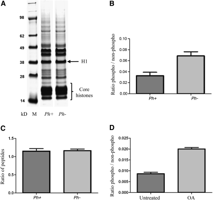
Figure 4.
Histone Extraction and Relative Levels of Phosphorylation in Histone H1 Peptides.
(A) Coomassie blue-stained histone extraction separated by 4 to 13% Bis-Tris gel in 2-(N-morpholino)-ethane-sulfonic acid-SDS running buffer. The locations of the linker histone H1 and the core histone proteins are noted. M, molecular weight protein marker with masses in kD.
(B) Ratio of amount of phosphopeptide DAAVD(pT)PAAKPAK to amount of peptide DAAVDTPAAKPAK in seven biological replicates of wheat–rye Ph1+ samples and nine biological replicates of Ph1− samples.
(C) Ratio of amount of two regularly observed peptides from histone H1 (SGSTIAIGK and ILLTQIK) in three biological replicates of wheat–rye Ph1+ samples and three biological replicates of Ph1− samples.
(D) Ratio of amount of phosphopeptide DAAVD(pT)PAAKPAK to amount of peptide DAAVDTPAAKPAK in two biological replicates of wheat–rye Ph1+ samples untreated and treated with okadaic acid. Bars represent se from the mean. OA, okadaic acid.

