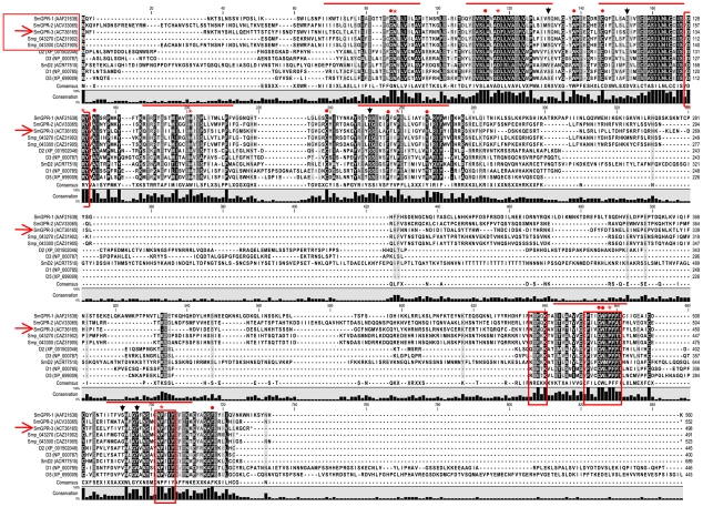
Figure 2. Sequence alignment of dopaminergic G protein-coupled receptors with Schistosoma mansoni SmGPR receptors.
A ClustalW alignment was performed using representative examples of vertebrate dopaminergic GPCRs (D1–D5), the S. mansoni dopamine D2-like receptor (SmD2) and several members of the SmGPR clade. SmGPR sequences are boxed (horizontal box) and SmGPR-3 is marked by an arrow. Receptor sequences are identified by their accession numbers (brackets). The positions of the predicted seven transmembrane domains are marked by horizontal lines and the invariant residue in each TM segment [37] is identified by an asterisk (*) Other conserved residues of functional relevance are marked by circles (•) and conserved motifs are boxed (vertical boxes). Residues discussed in this study, R2.64 (Arg96), D3.32 (Asp117), S5.42 (Ser198), T7.39 (Thr462) and Y7.43 (Tyr466) are identified by vertical arrows.

