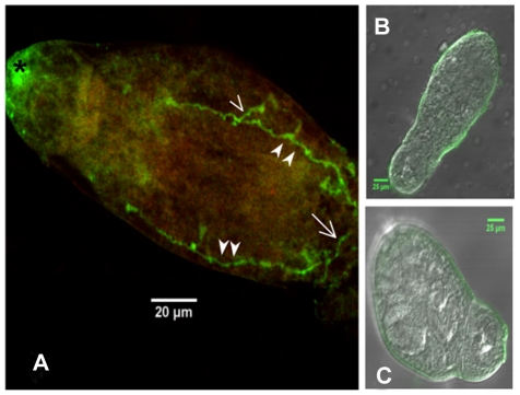
Figure 5. Immunolocalization of SmGPR-3 in larval Schistosoma mansoni.
S. mansoni cercaria were probed with affinity purified anti-SmGPR-3 antibody, followed by fluorescein isothiocyanate (FITC)-labelled secondary antibody. (A) Immunoreactivity (green) can be seen along the major longitudinal nerve cords (solid arrowheads) and in transverse commissures (open arrowhead), including the posterior transverse commissure near the base of the tail (open arrow). (B) No significant immunoreactivity was observed in negative controls probed with anti-SmGPR-3 antibody that was pre-adsorbed with peptide antigens or (C) controls probed with secondary antibody only. (*) non-specific labelling.

