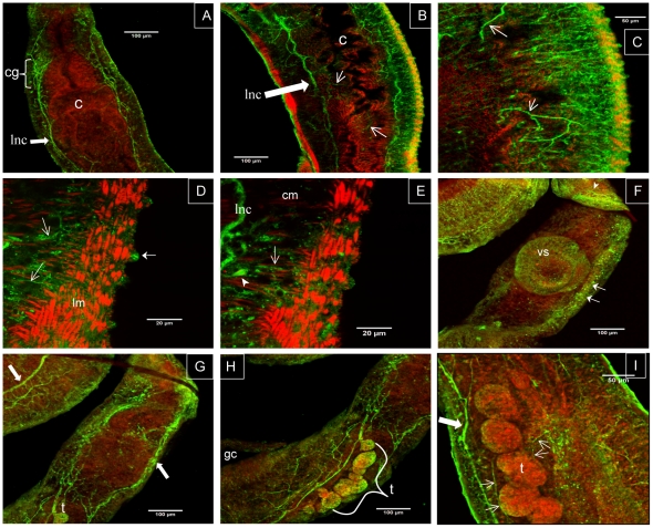
Figure 6. Immunolocalization of SmGPR-3 in adult Schistosoma mansoni.
Adult male worms were treated with affinity-purified anti-SmGPR-3 polyclonal IgG, followed by fluorescein isothiocyanate (FITC)-labelled secondary antibody (green). Animals were counterstained with tetramethylrhodamine B isothiocyanate (TRITC)-labelled phalloidin (red) to visualize the musculature of the body wall and digestive tract. (A) Strong SmGPR-3 immunoreactivity is visible in the region of the cerebral ganglia (cg) and along the main longitudinal nerve cords (lnc) of the CNS. (B) Green fluorescence can also be seen in peripheral nerve fibers innervating the caecum (open arrows). (C) Near the surface SmGPR-3 is strongly expressed in the peripheral innervation of the body wall muscles and the tegument. Numerous immunoreactive nerve fibers (open arrows), some varicose in appearance, are visible throughout this region. At higher magnification (D, E) we see SmGPR-3–expressing nerve fibers (open arrows) and cell bodies (solid arrowhead) innervating the body wall muscles, both circular muscle (cm) and typically spindle-shaped longitudinal muscle fibers (lm). SmGPR-3 immunoreactivity is seen in the tubercles of male worms (D, solid arrow), where it is probably associated with sensory nerve endings. (F) SmGPR-3 is expressed in the nerve plexus and small fibers of the ventral sucker. Extensive labelling of the submuscular nerve plexus can also be seen in this specimen (solid arrows). (G, H) A male worm showing strong labelling of major nerve cords (solid arrows) and the reproductive tract, including the testes (t) and associated nerves. (I) Fine SmGPR-3 immunoreactive nerve fibers (open arrows) innervate the testicular lobes of male worms. lnc, longitudinal nerve cords; cg, cerebral ganglia; c, caecum; lm, longitudinal muscle; cm, circular muscle; vs, ventral sucker; t, testes; gc, gynecophoral canal.

