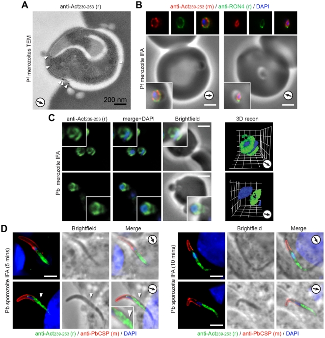
Figure 5. The spatial distribution of actin in invading merozoites and sporozoites.
A) Transmission electron micrograph with anti-Act239–253 (rabbit) immunogold labelling (arrowheads) of invading P. falciparum merozoite. Arrows show direction of invasion. B) Widefield IFA with deconvolution of invading P. falciparum merozoites labelled with mouse anti-Act239–253 (Red) or rabbit PfRON4 (Green) and DAPI (Blue). Scale bar = 2 µm. C) Widefield IFA with deconvolution of invading P. berghei merozoites labelled with rabbit anti-Act 239–253 (Green) and DAPI (Blue). Scale bar = 2 µm. Gamma settings were altered in 3D reconstruction. D) Widefield IFA with deconvolution of invading P. berghei sporozoites labelled with rabbit anti-Act239–253 (Green), anti-PbCSP (Red, exterior only) and DAPI (Blue). Scale bar = 5 µm, arrowhead shows presumed site of tight junction.

