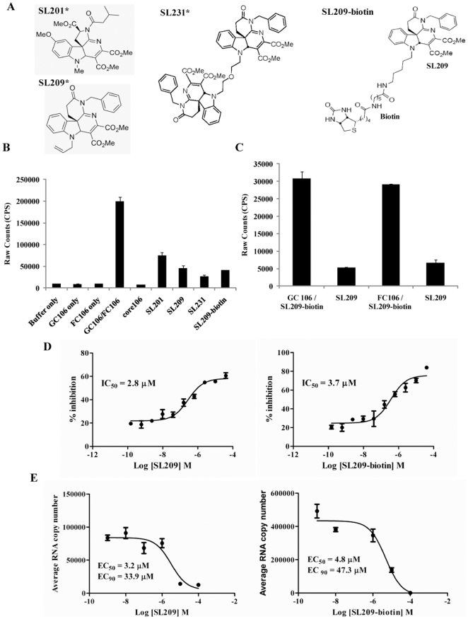
Figure 2. Inhibition of core dimerization and of virus production by SL209-biotin and evidence for direct binding to core protein.
In an AlphaScreen assay, glutathione-coated donor beads and anti-flag antibody coated acceptor beads (20 µg/ml each) were used to monitor the dimerization of GST- and Flag- tagged core106 proteins. GST-core106 (GC106) and Flag-core106 (FC106) were used at 150 nM each and core106 was used at 1 µM as a reference inhibitor. A: Structures of SL201, SL209, SL231, and SL209-biotin. SL201, is a 513 Da small molecule inhibitor originally identified to inhibit HCV core dimerization and virus production. SL209, is a SAR analogue of SL201. SL231, is a dimer of SL209. SL209-biotin is a biotinlyated derivative of SL209. “*” indicates that structures of SL201, SL209, and SL231 have been previously published in Wei et al 2009 [24], Strosberg et al 2010 [15], and Ni et al 2011 [23]. B: Levels of inhibition. Core106 inhibited 91% and small compounds inhibitors: SL201, SL209, SL231 and SL209-biotin used at 15 µM inhibited respectively 66%, 74%, 83%, and 75% of core dimerization. C: Direct binding to GST-core106 (GC106) or Flag-core106 (FC106). In a novel AlphaScreen format GC106/SL209-biotin and FC106/SL209-biotin were mixed in 1∶1000 ratio and incubated. Streptavidin donor beads and glutathione coated acceptor beads at 20 µg/ml were used in the detection of the binding. Free SL209 at 50 µM inhibited 83% of SL209-biotin binding to GC106 and 77% of SL209-biotin binding to FC106. D: Dose response analyses. Inhibition levels were analyzed in a dose response format. The compounds were dosed from 160 µM to 150 pM. The IC50 values for SL209 (right panel) and SL209-biotin (left panel) were calculated as 3.7 µM and 2.8 µM using GraphPad Prism. E: Inhibition of HCV production. Inhibition of HCV production in Huh-7.5 cells by SL209 and SL209-biotin was analyzed by adding serially diluted the compounds (individually) and virus onto naïve Huh-7.5 cells. The supernatants of the cells after 3 days of culture were removed from the initial culture and added to naïve cells cultured for another 3 days. RNA was purified from lysed cells and analyzed by Real-Time RT-PCR. EC50 values were calculated to be 3.2 µM and 4.8 µM for SL209 (right panel) and SL209-biotin (left panel), respectively. EC90 values were calculated to be 33.9 µM and 47.3 µM for SL209 (right panel) and SL209-biotin (left panel), respectively.

