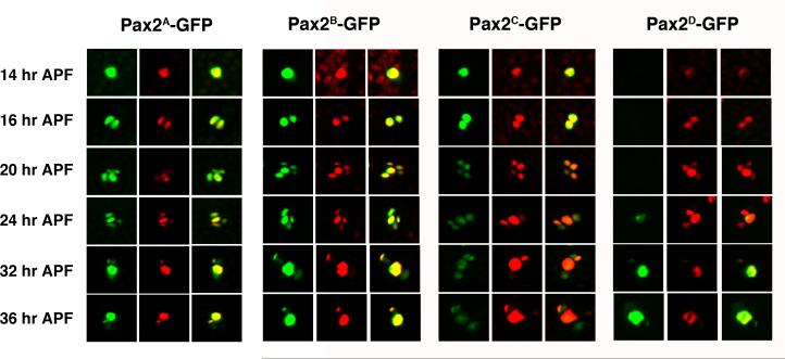
Figure 2. GFP reporter expression driven by different es organ enhancer regions.
Pupal nota from Pax2A-GFP, Pax2B-GFP, Pax2C-GFP and Pax2D-GFP reporter lines were dissected at 14, 16, 20, 24, 32 and 36 hours APF and stained with an anti-D-Pax2 antiserum. For each line and time point, the left hand panel shows GFP (green), the center panel shows D-Pax2 (red) and right hand panel is the merged image. Each panel shows a single es organ position in the developing microchaete field.

