
Fig. 1.
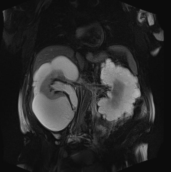
Coronal T2-weighted magnetic resonance imaging shows bilateral renal multilocular hyperintense subcapsular lesions. These findings are typical of renal lymphangiomatosis.
Official websites use .gov
A
.gov website belongs to an official
government organization in the United States.
Secure .gov websites use HTTPS
A lock (
) or https:// means you've safely
connected to the .gov website. Share sensitive
information only on official, secure websites.
Coronal T2-weighted magnetic resonance imaging shows bilateral renal multilocular hyperintense subcapsular lesions. These findings are typical of renal lymphangiomatosis.