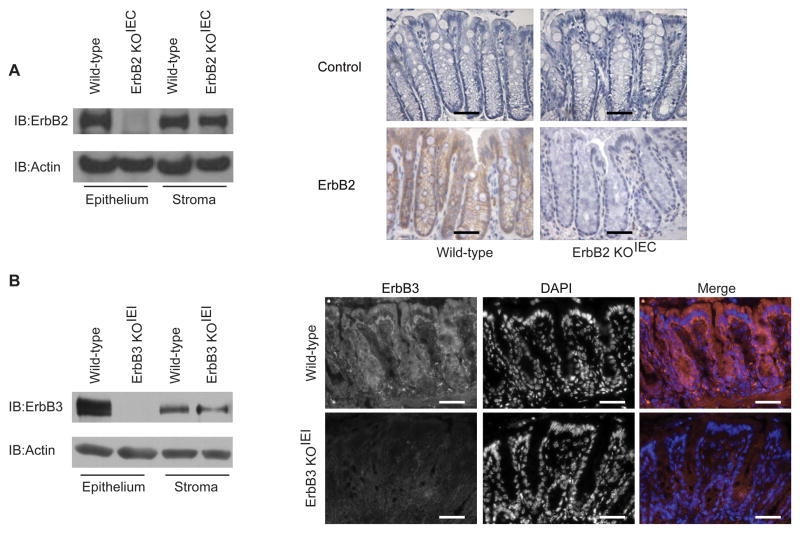
Figure 1. Deletion of colon epithelial ErbB2 or ErbB3 expression in ErbB2 KOIEC and ErbB3 KOIEI mice.
Epithelial and stromal fractions were isolated from colons of the indicated mice. (A and B) Western blot and immunohistochemical analysis with antibodies against ErbB2 (A) or ErbB3 (B). Omission of primary antibody was used as a control for immunohistochemistry. Actin expression was used as loading control. Brown indicates ErbB2 staining. Red indicates ErbB3 staining, blue indicates DAPI staining. Scale bars, 50 μm. These results are representative of independent analyses of at least three different mice in each group.

