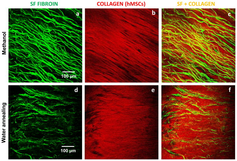
Figure 10.
Multi-photon microscopy evaluation of the lamellar 3D scaffolds treated with methanol and water annealing and after the construct was cultured up to 3 weeks. In green is the two-photon excited fluorescence image of the silk scaffolds (a) treated with methanol and (b) water annealed. In red is the SHG signal from the deposited collagen for the respective treatments (c,d).

