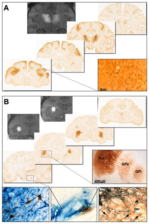
Fig. 1. Prediction of vector distribution by real-time MR imaging.
Co-infusion of gadoteridol and AAV2-GDNF allows real-time monitoring of distribution at the site of infusion that is highly predictive of the area of GDNF expression. Representative MR images and immunohistochemical staining for GDNF after bilateral infusion into the NHP (A) thalamus or (B) putamen (2 sites per hemisphere). MR imaging immediately after completion of the infusions showed that the gadoteridol tracer was confined to the target structures with no leakage or backflow along the cannula tracts. The distribution of GDNF in the thalamus and putamen correlated exactly with the MR signal. In addition, anterograde transport of AAV2-GDNF to secondary brain regions was observed with GDNF present in multiple regions of the cortex after infusion into the thalamus, and basal ganglia nuclei after infusion into the putamen. Dual staining for GDNF (brown) and tyrosine hydroxylase (blue) shows some overlap between GDNF expression in the substantia nigra and TH-positive neurons in the pars compacta. However, many GDNF-positive neurons (arrows) were found in the TH-negative SNr. Abbreviations: GPe: Globus Pallidus external; GPi: Globus Pallidus internal; Put: Putamen; SNc: Substantia nigra pars compacta; SNr: Substantia nigra pars reticulata; VTA: ventral tegmental area.

