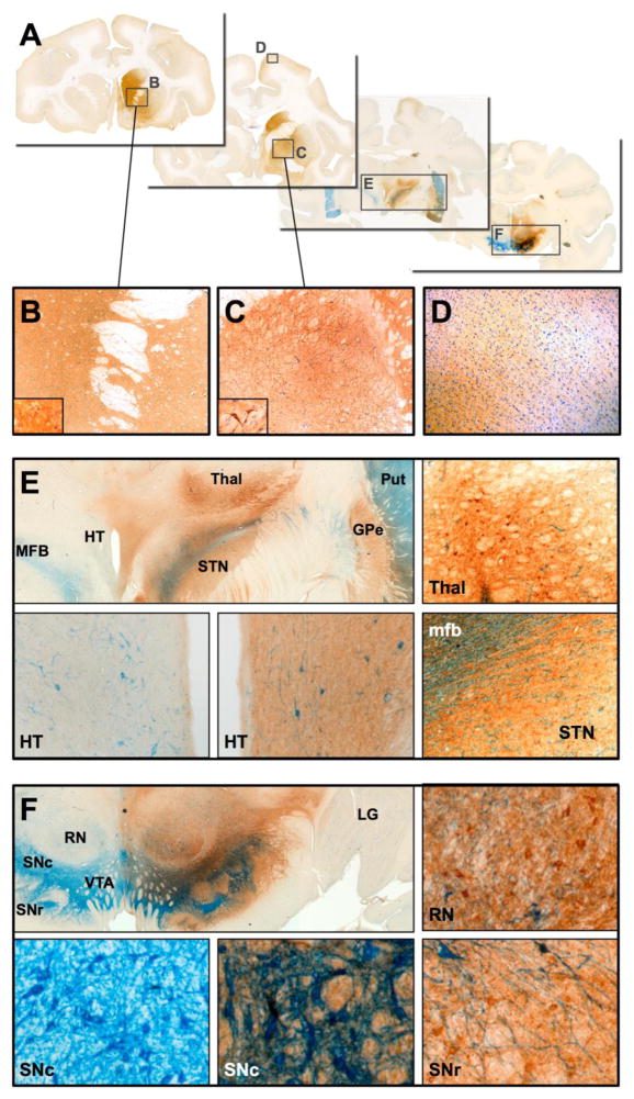
Fig. 3. Distribution of GDNF after delivery of AAV2-GDNF to the midbrain.
(A) Direct infusion of AAV2-GDNF into the substantia nigra of aged NHP resulted in extensive GDNF expression in many distal areas of the primate brain, including (B) caudate nucleus, medial putamen, (C) globus pallidus and (D) cerebral cortex. GDNF-positive neurons and fibers (E) were also found in the ventral thalamus, hypothalamus and subthalamic nucleus, none of which were directly targeted during AAV2-GDNF infusion. Within the targeted midbrain, extensive GDNF expression was present (F) in the substantia nigra, ventral tegmental area and red nucleus. No GDNF was observed in the contralateral hemisphere. IHC staining: tyrosine hydroxylase (blue), GDNF (brown).

