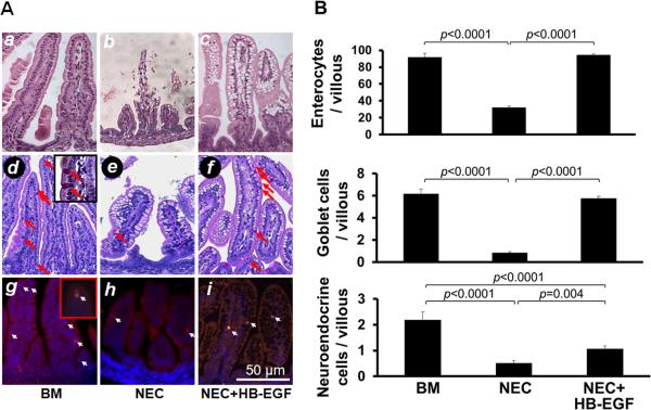
Figure 1.
HB-EGF protects enterocytes, goblet cells and neuroendocrine cells from injury due to experimental NEC. (A) Representative photomicrographs of rat jejunum. a-c) H&E stained sections showing absorptive enterocytes; d-f) PAS staining showing goblet cells (red arrows); and g-i) chromogranin A immunostaining showing neuroendocrine cells (white arrows). Panels a, d and g are from pups that were breast fed; panels b, e and h are from pups subjected to experimental NEC; panels c, f and i are from pups subjected to experimental NEC but with HB-EGF added to the feeds. (B) Quantification of enterocytes, goblet cells and neuroendocrine cells. BM, pups that were breast fed by surrogate mothers; NEC, pups exposed to experimental NEC; NEC+HB-EGF, pups exposed to experimental NEC that were treated with HB-EGF added to the feeds. Values represent mean ± SEM. One-way ANOVA with Tukey-Kramer pair-wise comparison test.

