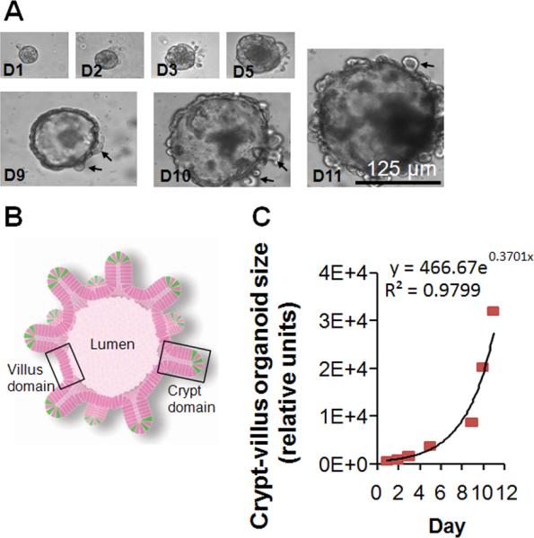
Figure 3.

Ex vivo crypt-villous organoid cultures. (A) A representative mouse crypt cultured ex vivo in gelmatrix that grew into a crypt-villous organoid with budding crypts (black arrows) extending from the surface at days 1-11. (B) Organoid illustration showing lumen, villus domain, crypt domain, and ISCs in green (from Sato et al.28) (C) Exponential growth of crypt-villous organoids cultured with R-spondin 1, Noggin and HB-EGF (50 ng/ml).
