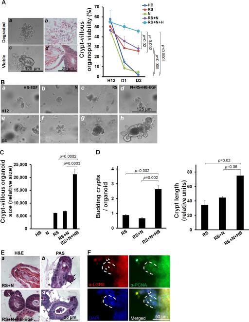
Figure 4.
HB-EGF protects crypt-villous organoids from hypoxia. (A) Representative photomicrographs of viable and degraded organoids as visualized by phase contrast microscopy (panels a,c) and H&E staining (panels b,d). Percent of viable organoids with different culture additives at 12 hours (H12), day 1 (D1) and day 2 (D2). (B) Representative crypt-villous organoids cultured for 12 hours or 4 days in medium containing: a, e) HB-EGF; b, f) Noggin; c, g) R-Spondin 1; d, h) Noggin + R-spondin 1 + HB-EGF. Panels a-d: 12 hours; panels e-h: day 4. (C) Size of crypt-villous organoids cultured with different additives on day 4. (D) Budding crypts/crypt-villous organoid cultured with different additives on day 4 and budding crypt length cultured with different additives on day 4. (E) Crypt-villous organoids (day 4) cultured with R-spondin and Noggin, in the absence (panels a, b) or presence (panels c,d) of HB-EGF (50 ng/ml). Panels a,c: H&E staining; panels b,d: PAS staining; white arrows, Paneth cells; black arrows, goblet cells. (F) Crypt-villous organoids (day 4) cultured with R-spondin and Noggin and HB-EGF (50 ng/ml), and immunostained with anti-LGR5 (Cy3, red) and anti-PCNA (Cy2, green), to identify ISCs and TA progenitor cells, respectively. The white dashed lines indicate budding crypts and the white arrows indicate ISCs at the base of the crypts. In A-E: RS, R-spondin 1; N, Noggin; HB, HB-EGF. In B-C: values represent mean ± SEM. B (right panel), Two-way ANOVA; B (left panel) and C, one-way ANOVA with Tukey-Kramer pair-wise comparison test.

