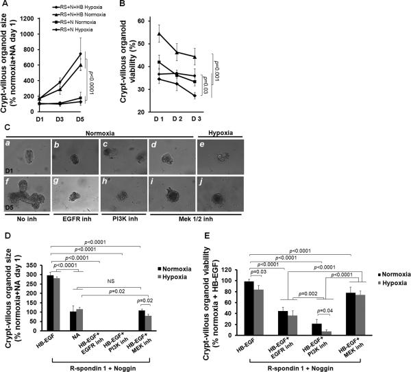
Figure 5.
HB-EGF promotes crypt proliferation under hypoxic stress via EGFR activation and PI3K/Akt and MEK1/2 pathways. (A) Organoid size after exposure to hypoxia for 60 min followed by culture for 1-5 days in the presence of R-spondin 1 and Noggin, with or without HB-EGF (50 ng/ml). (B) Percent of viable organoids after exposure to hypoxia for 60 min followed by culture for 1-3 days in the presence of R-spondin 1 and Noggin, with or without HB-EGF (50 ng/ml). (C) Representative photomicrographs of crypt-villous organoids cultured in the presence of R-spondin 1, Noggin and HB-EGF (50 ng/ml), plus the addition of: a,f) no inhibitors; b,g) AG 1487 (EGFR inhibitor); c,h) LY294002 (PI3K inhibitor); d,e,i,j) PD98059 (MEK1/2 inhibitor). Panels a-e represent day 1 of culture; panels f-j represent day 5 of culture. The organoids in panels e and j were exposed to hypoxia (100% nitrogen for 60 min); the organoids in all other panels were exposed to normoxia. (D) Organoid size on day 5 of culture, in the presence of R-spondin 1 and Noggin and HB-EGF, with or without inhibitors, upon exposure to hypoxia or normoxia. (E) Quantification of viable organoids on day 1 of culture, in the presence of R-spondin 1 and Noggin and HB-EGF, with or without inhibitors, upon exposure to hypoxia or normoxia. NA, medium containing R-spondin 1 and Noggin with no addition of HB-EGF; N, normoxia; H, hypoxia; D1, Day 1 of culture; D2, Day 2; D3, Day 3, D5, Day 5. Values represent mean ± SEM. Two-way ANOVA with Tukey-Kramer pair-wise comparison test.

