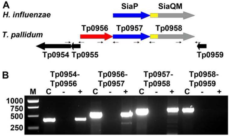
Figure 1. Transcriptional linkage of the putative treponemal TRAP operon.
(A) The genomic organizations of TRAP systems from H. influenzae (upper) and T. pallidum (lower). The genes for the P components are colored blue, those for the Q components are yellow, and those for the M components are gray. These latter two components are fused in the examples shown, but not in all TRAP-Ts. (B) Evidence of transcriptional linkage of Tp0956 and Tp0957. RT PCR was performed on T. pallidum RNA using primer pairs (small arrows) listed in Table S1, specific for either the intergenic regions of listed gene pairs or the entire tp0955 gene (pseudo gene). The lanes are: Lane M, DNA molecular weight markers; Lane C, PCR reaction with indicated primer pair served as positive control using T. pallidum genomic DNA as template in place of cDNA; Lane ‘-’, PCR reaction with indicated primer pair using RNA as template (lacking RT), which served as a negative control for DNA contamination; Lane ‘+’, RT PCR products with indicated primer pairs.

