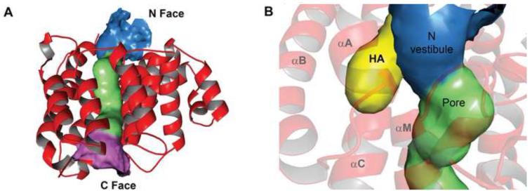
Figure 3. Pore characteristics of Tp0956.
(A) A ribbons representation of the protein with a surface representation of the pore. The N, middle, and C portions of the pore are colored blue, green, and pink, respectively. (B) The hydrophobic antechamber (HA). A different view of the pore than that in (A) is shown, and the HA is depicted as a yellow surface. Secondary structure is shown semi-transparently for clarity, and the α-helices that contribute residues to the HA are labeled. The HA surface was calculated with CAVER 2.0 71.

