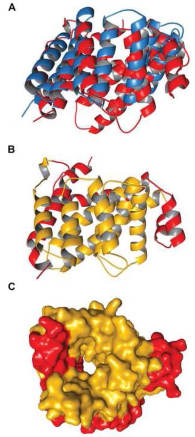
Figure 4. The cTPR motifs of Tp0956.
(A) A superposition of Tp0956 (red) and DrR162B (blue). (B) The positions of the cTPR motifs. The motifs are colored gold, while the remainder of the protein is red. (C) The contributions of the cTPR motifs to the pore of Tp0956. A surface representation is shown, with the protein colored as in (B).

