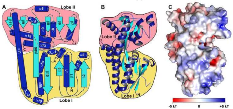
Figure 7. The topology, structure, and surface features of Tp0957.
(A) A topology diagram of Tp0957. β-strands are shown as cyan arrows, while α-helices are depicted as blue cylinders and 310 helices are shown as ovals. The β-strands and α-helices are shown to scale, but the connecting regions and 310 helices are not. All elements are labeled with the names that will be used throughout the text. Lobe I is outlined in yellow, Lobe II in pink. The amino-and carboxyl-termini are marked with “N” and “C”, respectively. (B) A ribbons-style drawing of the structure of Tp0957. The secondary structure and lobes are color-coded as in part (A). (C) The surface electrostatic features of Tp0957. The electrostatic potential is color-coded on the surface of Tp0957. A key to the coloring scale is shown. The orientation of Tp0957 is identical to that in part (B).

