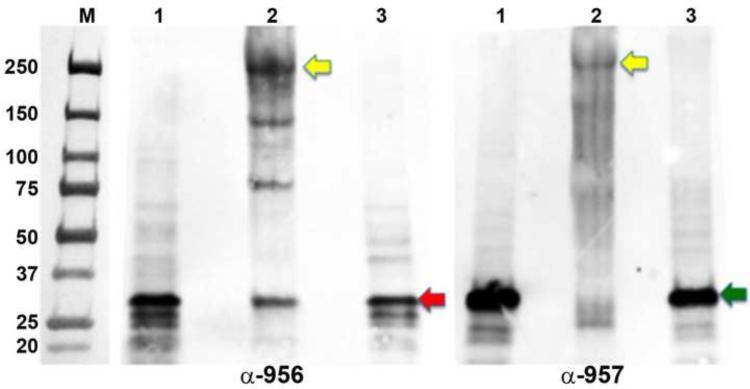
Figure 9. In vivo formaldehyde cross-linkage of Tp0956 and Tp0957.
Samples were resolved in two identical gels and then electrotransferred to two separate membranes. The membranes were individually probed with either an anti-Tp0956 (α-Tp0956; left panel) or an anti-Tp0957 (α-Tp0957; right panel) antibody and visualized by an enhanced chemiluminescent detection using an anti-goat IgG-HRP. Lane 1 shows T. pallidum not exposed to the formaldehyde; lane 2, formaldehyde cross-linked treponemes, and lane 3, boiled treponemes to break the cross-links prior to SDS PAGE. The yellow arrows represent the putative positions of the cross-linked hetero-hexameric complex, while the red and green arrows denote the positions of monomeric Tp0956 and Tp0957, respectively. The positions of pre-stained molecular-weight standards (M; values are in kDa) are indicated in the image of the blot.

