
Figure 1.
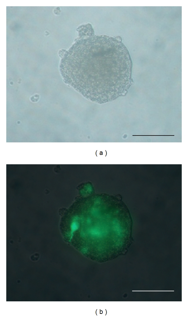
LV-GFP-labeled secondary neurosphere from adult pig SVZ, just before transplantation and 3 days after in vitro infection with the lentiviral vector of GFP, as observed under a photonic microscope with natural light (a) or GFP fluorescence (b). Scale bars: 50 μm.