
Figure 3.
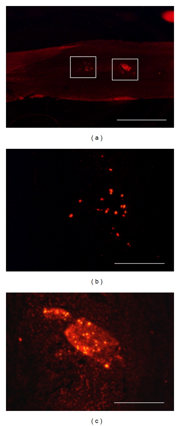
Localization of transplanted BrdU-labeled neurospheres in the bridged nerve at 8 days after lesion, by postmortem immunohistochemistry. BrdU immunoreactivity is revealed by red Alexa-594 fluorescence on longitudinal postfixed graft sections at low (a) and high (b, c) magnifications. Enlarged fields (b, c) are localized as white forms in (a). Transplanted neurosphere cells are found either sparsed (b) or clustered (c) inside the venous bridge. Scale bars: 0.5 mm (a), 100 μm (b, c).