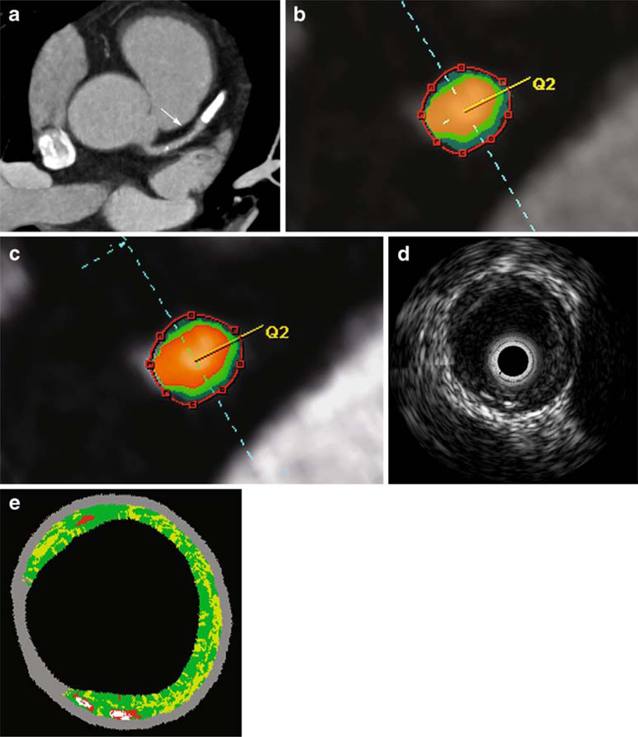
Fig. 6.
Virtual plaque histology in DSCT and IVUS. In an axial MIP, the arrow indicates a non-calcified plaque in segment 6 of the LAD (a). Cross-sectional images of the vessel with color coding are presented for visual adjustment of the maps (b) or HU-based segmentation (c) as well as for grey-scale IVUS (d) and IVUS-VH (e). For IVUS-VH, red indicates necrotic core, light green the fatty-fibrous, dark green the fibrous and white the calcified plaque. In this lesion, total plaque volumes with visual CT evaluation, HU-based analysis or IVUS were 66.6 mm3, 55.8 mm3 or 53.1 mm3 respectively

