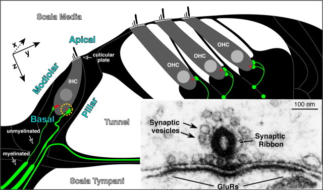
Figure 1.
Schematic cross section through the cochlear epithelium showing the unmyelinated afferent terminals (green) on IHCs and OHCs and the presynaptic ribbons at each synapse (red). The modiolar and pillar side of the IHC, its apical versus basal pole, and the position of the cuticular plate are indicated. Inset shows an electron micrograph (Liberman, 1980b) of the presynaptic ribbon in an IHC, its halo of synaptic vesicles, and the postsynaptic membrane density on the terminal swelling, where GluRs are located. Approximate orientation of x-, y-, and z-planes in the subsequent confocal images is shown: images are acquired from epithelial whole mounts, viewed from the scala media surface, thus the x-axis runs into, and out of, the plane of the schematic.

