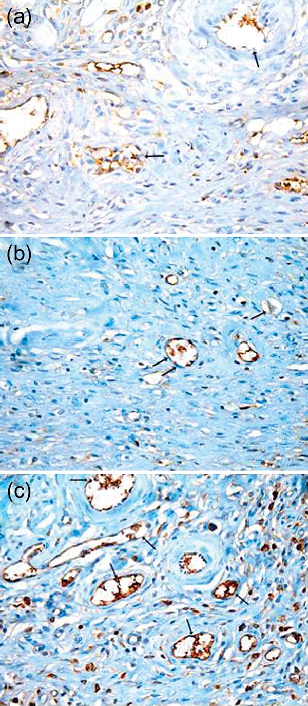
Figure 3:

(a) Photomicrograph of a skin section from the control group showing normal vasculature in the dermis of the wound area; however, the reaction is mostly negative in the endothelial cells (arrow). (b) Photomicrograph of a skin section from the DC 2 week group showing an area of the dermis with decreased number of blood vessels. The reaction in the endothelial cells is mostly negative (arrow). (c) Photomicrograph of a skin section from the St 2 week group showing an area of the dermis with increased number of blood vessels which are mostly functioning and contains RBCs (arrow). There is a strong positive reaction in the endothelial cells lining the vessels. (Immunostaining for CD31 anti-body ×400.)
