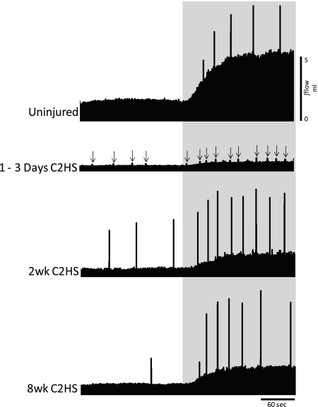
Fig. 2.
Representative examples of tidal volume (VT) during spontaneous breathing at baseline and hypercapnic respiratory challenge. Images show the integrated airflow signals (∫flow) from a control (uninjured) rat and rats studied 1–3 days, 2 wk, and 8 wk following C2HS injury. Shaded area represents the hypercapnic challenge (7% inspired CO2). Spikes in the record are augmented breaths (ABs; see text); the ABs were harder to detect at 1–3 days post-C2HS and are indicated by arrows. C2HS resulted in decreased VT during both baseline and hypercapnic challenge at all postinjury time points. In addition, a reduction in the volume of ABs was observed following C2HS. Scaling is identical in all panels.

