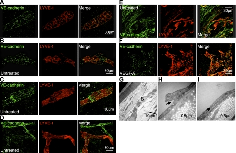
Figure 5.
Lymphatic endothelial junctions in cornea and conjunctiva. A–F) Confocal images of double-stained corneal flatmounts for LYVE-1 (red) and VE-cadherin (green) in untreated corneas (A–C), untreated conjunctiva (D, E), and VEGF-A-stimulated corneas (F; 200 ng, d 6) of C57BL/6 mice. G) Electron-microscopic images of untreated corneas. L, lymphatics; B, blood vessels. H, I) Arrows indicate overlapping valve-like lymphatic endothelial endings.

