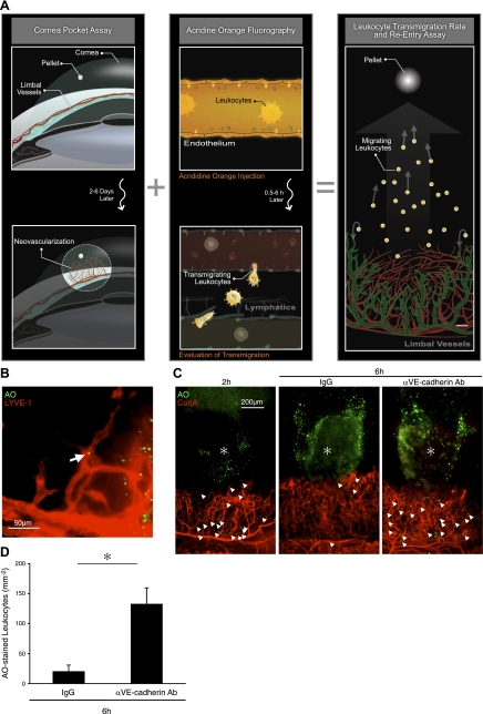
Figure 7.
VE-cadherin-dependent reentry of transmigrated leukocytes into lymphatics. A) Schematic of our new imaging technique for visualization of transmigrated leukocytes. B) AO+ leukocytes in FGF-2-induced lymphangiogenic vessels of C57BL/6 mice with LYVE-1 staining, 2 h after AO injection, 4 d after pellet implantation. C) AO+ leukocytes and Con A+ angiogenic vessels in FGF-2-implanted corneas, 2 and 6 h after AO injection, 4 d after pellet implantation. Topical anti-VE-cadherin Ab or IgG treatments, 2 h after AO injection. D) Quantitation of the number of AO+ leukocytes in angiogenic areas of FGF-2-implanted corneas, 6 h after AO injection. (n=3–4). *P < 0.05.

