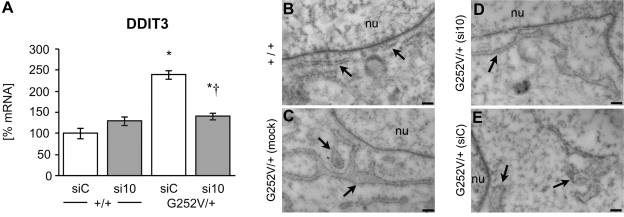
Figure 5.
Effect of allele-specific RNAi on the ER UPR in human fibroblast tissue culture. A) Amount of DDIT3 mRNA was analyzed in wild-type and COL3A1G252V/+ fibroblasts by real-time RT PCR. Fibroblasts were treated with 100 nM siC (control) or si10 siRNA for 2 d. Data are averagse ± se of 3 biological and 4 technical replicates. Results were analyzed by the 2−ΔΔCT method normalized to GAPDH. Result of a nonsilencing control was set as 100%. *P < 0.05 vs. WT control; †P < 0.05 vs. COL3A1G252V/+ control. B–E) Analysis of the morphology of the ER of cultured primary fibroblasts by transmission electron microscopy. Fibroblasts from cell cultures were collected after trypsin/EDTA treatment and embedded in LRwhite for ultrathin sectioning. In normal fibroblasts (B), the ER close to the nucleus appeared as long and regular tubes, whereas the ER of fibroblasts with the Col3A1G252V/+ mutation (C) showed a broadening of the tubes with several expansions. Treatment of the mutated cells with siRNA 10 (D) resulted in changes of the ER structure toward the typical morphology of normal fibroblasts. In contrast, the morphology of the ER was not affected when a nonsilencing siRNA was applied (E). Tubes of the ER were still enlarged. Arrows indicate ER. nu, nucleus. Scale bars = 200 nm.

