
Fig. 3.
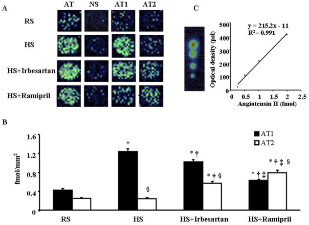
(3-[125I]iodotyrosyl4,Sar1-Ile8)Angiotensin II binding in LV sections. A: Pseudocolour images of LV sections from mice subjected to regular salt (RS) or high salt (HS) diet and HS mice treated with irbesartan (HS+Irbesartan) or ramipril (HS+Ramipril) for 8 weeks. LV sections were incubated with (3-[125I]iodotyrosyl4,Sar1-Ile8)Angiotensin II alone (AT: total angiotensin II binding) or in the presence of unlabelled angiotensin II, PD-123319, or losartan which showed the nonspecific (NS), AT1 and AT2 binding, respectively. B: Mean values of AT1 and AT2 densities in the left ventricle (n=5 in each group). C: Typical calibration curve and corresponding quantification. * p<0.05 versus RS mice, † p<0.05 versus HS mice, ‡ p<0.05 versus HS+Irbesartan and § p<0.05 versus corresponding AT1 value.