
Figure 3. Functional overexpression and knockdown of βIII-tubulin modulates the androgen-independent prostate cancer cells sensitivity to docetaxel.
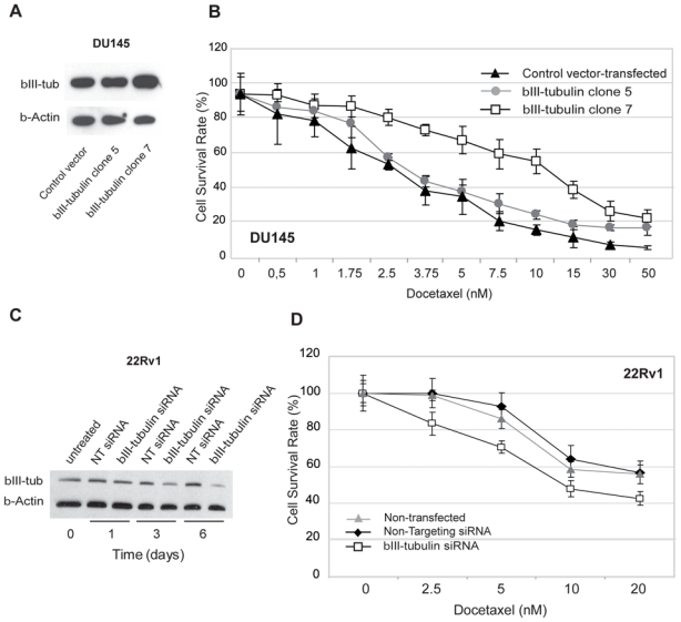
A. Differential levels of βIII-tubulin protein between control vector-transfected and βIII-tubulin transfected (clone 5 and 7) DU145 cells: Western blot analysis showed that. The βIII-tubulin clone 5 expressed a similar level of βIII-tubulin compared with control vector-transfected cells, whereas the βIII-tubulin clone 7 showed a higher level of βIII-tubulin expression.
B. Dose response curve assessing the effect of βIII-tubulin overexpression in androgen-independent DU145 cells. Cell viability assays showed that clone 7 was significantly more resistant to docetaxel treatment relative to βIII-tubulin clone 5 and control vector-transfected DU 145 cells. Points, mean; bars, SEM. NT: non targeting.
C. Western blot confirmation of βIII-tubulin knockdown in 22Rv1 cells after siRNA transfection. The βIII-tubulin protein level was decreased 72 hours after transfection and lasted at least 6 days after a single transfection.
D. Dose response curve assessing the effect of βIII-tubulin silencing in androgen-independent 22Rv1 cells. Cell viability assays showed that βIII-tubulin siRNA-transfected 22Rv1 cells were significantly more sensitive to docetaxel treatment relative to non targeting siRNA-transfected and non-transfected 22Rv1 cells. Points, mean; bars, SEM.