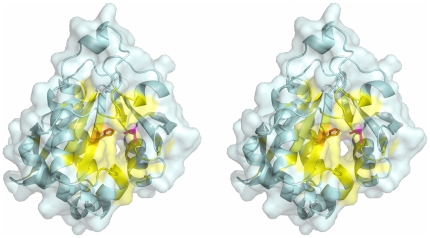
Figure 8. High-quality functional site prediction with text evidence for PDB entry 1YK3 [40].
The protein is displayed in wall-eyed stereo pairs as a light blue ribbon with a semitransparent surface. The predicted functional site from structure-based analysis is colored yellow, and the predicted residues mentioned in the abstract of the primary reference are rendered as sticks colored orange (His130) and magenta (Asp168).

