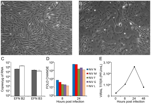
Figure 1. Primary human endothelial cells are highly permissive to NiV-infection.
A, Mock-infected and B, NiV-infected HUVECs (MOI = 1) observed 24 h after infection show extensively developed syncytium formation. C, RT-qPCR analysis of the expression of NiV receptors ephrinB2 (EFNB2) and ephrinB3 (EFNB3) in HUVECs (black bars) and U373 astroglioma cells (white bars). D, Production of RNA specific for NiV genes: nucleoprotein (N), matrix (M), fusion protein (F), glycoprotein (G) and polymerase (L) in HUVECs during infection, determined by RT-qPCR. Fold change is relative to the number of copies of viral mRNAs 8 h or 24 h post infection compared to the number of copies obtained after 1 h of contact with the virus. E, Production of infectious NiV particles during the course of infection; supernatants taken at different time points were titrated on a Vero cell monolayer.

